|
Mosaic-like colorations in pigeons
At the time of the early reports on mosaics by Cole and Hollander,
it was the name of a phenotype whose possible different causes were
speculated about. The terms mosaic, chimera and somatic mutations
stood side by side, mosaic as a generic term. 'Somatic Mosaics' is
the headline of an article by Cole and Hollander, submitted in 1939
and published in 1940 in the scientific journal 'Genetics'.
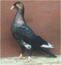 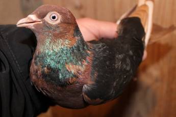
Fig. 1: Highflier-hens with mosaic-like contrasting color areas from
the own loft at the left and at Rudolf Beneke, at the right.
With 'Bipaternity' in the title of an article in the 'Journal of
Heredity' in 1949, Hollander expressed his suspicion about the cause
of some mosaic-like colored pigeons, in which other explanatory
approaches could not apply due to their potential descent, or only
when several unlikely events interacted. With advances in molecular
genetics, other explanations came to the fore. The 'mosaic or
chimeric effects' summarized by Hollander / Cole in 1940 were
divided into chimera and mosaic according to the suspected causes.
What they have in common is that they have genetically different
cell populations. In the case of chimeras, they originate
from the fusion of two or more fertilized egg cells (zygotes); in
the case of mosaics, they originate from a single zygote,
https://www.britannica.com/science/chimera-genetics,
https://www.embryology.ch/vet/de/kchromaber/klinik02.html
How do we get genetically different cells into a fertilized egg
cell? Mutations and deletions are possible. Somatic mutations
only set in after the embryo has formed. From then on, the DNA
sequence of body cells is changed. These changes can only be
reproduced in parts. The change is limited to the body cells and is
not inherited. Theoretically, the distinctions are clear, but
difficult in practice, as publications from human medicine show (e.
g. Boklage 2006). In the case of pigeons, there is usually little
evidence of potential parentage.
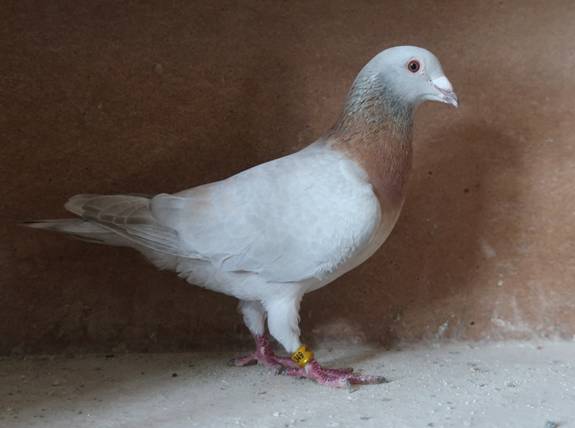
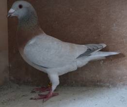
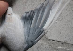
Fig. 2: Cock with mosaic-like contrasting color areas
In the case of the cock from the own loft shown, it can be narrowed
down. The father is a pure, homozygous rubella. The mother is
hemizygous, a dilute frosty rubella check. Typical for such hens -
with a genetically black basic color - the yellow color in the
phenotype.
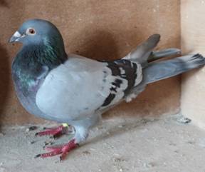 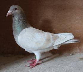
Fig. 3: Parents of the cock in Fig. 2. Homozygous rubella cock and
hemizygous dilute frosty rubella hen.
Outwardly, the young cocks mostly show the plumage coloration
expected with this genetic makeup. Compared to most homozygous
rubella, heterozygous frosty cocks the color is weakened in a
greater degree and thus similar to hemizygous frosty rubella hens
(Fig. 5 at the left) and not far away from some homozygous frosty
that were classified as heterozygous rubella. Homozygous Frosty
Rubella cocks are light silver-gray with translucent bars or checks.
At the cock in Fig. 2 we see mosaic-like deviations on one side in
several primaries, thumb springs and hand covers. Here he shows the
coloring of pure, non-diluted Rubella.
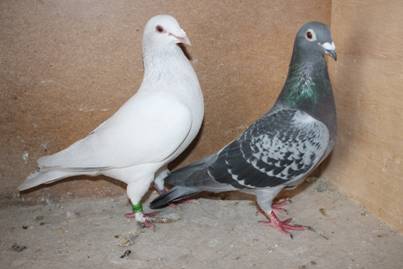
Fig. 4: Grandparents of the cock in Fig. 2 from the mother side:
Homozygous Frosty-Rubella cock, black color base, bar pattern,
homozygous frosty and rubella, heterozygous dilution, Non-Spread. At
the right the blue check grandmother
The potential genetic makeup based on the family tree: The son
should have inherited the frosty gene, rubella and the dilution
factor on the maternal sex chromosome. On the paternal side,
non-frosty, rubella and non-dilution are to be expected on the
chromosome at the gene locations mentioned. The majority of the
plumage corresponds to the expectations of this genetic makeup. The
relatively strong lightening in this cock and the yellowish tinge,
especially in the neck area, are probably due to the fact that it is
also heterozygous for dilution on the maternal side.
With the known descent one can mentally pursue the potential
inheritance processes. In the mosaic-like deviations, the maternal
expected frosty gene does not have an effect. The gene for this area
of the body seems to be lost in the son's inheritance. It is not
due to the father and cannot be explained by another paternity
(cross-fertilization) from the breeding group. Another pure-bred
rubella cock would be irrelevant. A wild-type bluebird and the
pure-bred Frosty Rubella grandfather are also ruled out, as they
could not have resulted in the appearance in combination with the
mother's genes. Bipaternity as an explanation for appearance
falls out for the same reason.
In chimeras, two fertilized egg cells fuse. On the maternal
side, the hen can only form the type frosty, rubella, dilution on
the sex chromosome. Combined with the genes on the sex chromosome of
potential fathers, no zygote for the observed phenotype can result
from this. Mosaics arise from a fertilized egg cell, whereby
mutations are one possibility of genetically different cells. On the
maternal side, Frosty could have mutated back to the wild type. If
this occurs after the embryo has formed, only certain body cells are
affected (somatic mutations or somatic mosaic). In
combination with the unchanged paternal chromosome, this can result
in a mosaic pattern, as shown.
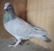 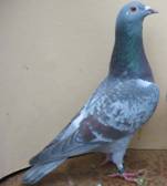 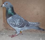
Fig. 5: On the effect of Frosty at a Rubella base: At the left a
hemizygous frosty-rubella hen, in the centre a hemizygous rubella
hen. Source: Sell, Critical Issues in Pigeon Breeding Part III). At
the right a homozygous rubella check cock
Other examples of mosaic-like colored pigeons suggest chimera as the
cause. Still others are difficult to reconcile with both theories or
require a large number of coincidental peculiarities at the same
time. For the classic analysis by looking at the phenotypes, there
is not only the uncertainty about the ancestry, but also that little
or nothing is known about many interactions of color factors. This
also applies to the interaction of factors that are not allelically
assessed as recessive. They are recessive in mating with the wild
type, but cause clear color changes in certain gene constellations
even when heterozygous. http://www.taubensell.de/extreme_sexual_dimorphism_in_the.htm
 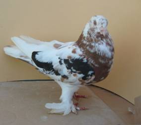
Fig. 6: Dark color
patches in a cock with the stipper gene and in an Uzbek Flying
Tumbler, 'Tschinny', from the own loft. Tschinnies are self red in
the juvenile plumage. Such patches are common with these stains and
are also reported from other lofts (Source: Sell, Pigeon Genetics)
In the case of pigeons, there seem to be no meaningful empirical
studies on the chromosomal differences between chimeras and mosaics
and investigations of the body cells.
The relationship between the extent and the distribution of the
mosaic areas with the potential cause and the time of mutations in
supposed somatic mosaics has probably not yet been examined in
pigeons.
The general difficulties of differentiation in specific cases are
known from human genetics. As Boklage (2006) states, mosaic
formation is not a problem in terms of the theoretical definition,
but in everyday clinical practice and in diagnostics.
Literature:
Boklage, Charles E. (2006) Embryogenesis of chimera, twins and
anterior midline asymmetries, Human Reproduction Vol. 21, No. 3, pp.
579-591.
Hollander, W.F. (1949), Bipaternity in Pigeons, Journal of Heredity,
Vol. XI., No. 10, pp.271-277.
Hollander, W.F., und Leon J. Cole (1940), Somatic Mosaics in the
Domestic Pigeon, Genetics Vol. 25, pp. 16-40.
http://www.taubensell.de/extreme_sexual_dimorphism_in_the.htm
http://www.taubensell.de/extremer_geschlechtsdimorphismus.htm
https://www.britannica.com/science/chimera-genetics
Sell, Axel, Pigeon Genetics. Applied Genetics in the Domestic
Pigeon, Achim 2012.
Sell, Axel,
Taubenzucht. Möglichkeiten und Grenzen züchterischer Gestaltung,
Achim 2019.
Universities of Fribourg, Lausanne and Bern (Switzerland), Online
course in embryology for medicine students, developed by the
Universities of Fribourg, Lausanne and Bern with the support of
Swiss Virtual Campus, (on sight august 11, 2021)
https://www.embryology.ch/genericpages/credits.html
|